Because the prostate is a small organ located deep in the pelvis, traditional imaging methods have not been very successful in detecting prostate cancer. However, utilizing new generation 3T MRI scanners and special parameters, with interpretation of images by an experienced radiologist, greater than 97% accuracy can now be achieved in detecting clinically significant prostate cancer or excluding significant cancer.
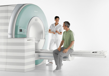
At Partners Imaging, we use a new generation Siemens Multiparametric (Mp) 3T MRI scanner with special protocols and computer aided detection software (CAD). I exclusively interpret all prostate MRI cases for Partners and have read thousands of exams.
If you are going to be scanned, shouldn’t you do it where it will give you the best chance for a diagnosis? A 3T MRI is the most powerful clinical magnet available. It provides twice the magnetic strength of the next lower strength magnet at 1.5T. For MR Imaging, signal strength is the most important factor.
The advantages of 3T MRI over 1.5T or lower strength MRIs are:
a) Increased spatial resolution for improved anatomical detail.
b) Shorter scan times.
c) Increased number of high resolution images during dynamic contrast enhancement (DCE).
d) Spectroscopy & diffusion (parameters) are better at 3T because of increased signal.
e) An endo-rectal coil is usually not needed at 3T which can be uncomfortable and often results in scan artifact and distortion.
When you are scanned on our 3T MRI, high resolution anatomic T1 and T2 weighted images of the prostate are obtained and combined with at least 2 other parameters. The parameters include:
a) Dynamic Contrast Enhancement (DCE) – With an IV injection of FDA-approved Gadolinium, tumor vascularity is evaluated. Tumors “take up” and “wash out” contrast material quickly. The amount and type of enhancement is specific for tumor when correlated with T2 and DWI.
b) Diffusion Weighted Imaging (DWI) – Measures movement of water and tumor density. DWI has a high correlation with tumor grade or aggressiveness (Gleason score).
c) Magnetic Resonance Spectroscopy (MRS) – Measures chemicals or metabolites within cells. An increased Choline/Citrate ratio is specific for tumor.
High resolution anatomic T1/T2 imaging plus a) and b) above are used in the majority of cases for accurate detection of prostate cancer. In some cases, spectroscopy is recommended, especially in patients unable to receive IV contrast. Special post processing software improves image interpretation by providing quantitative analysis and tumor mapping. At Partners Imaging, all of this is provided on our dedicated multiparametric 3T MRI scanner for the most optimal exam.